Shoulder Tendon Anatomy / Shoulder Cartilage And Tendon Injuries My Doctor Online : Home » what is frozen shoulder » shoulder anatomy.. More precisely, it consists of several joints that work together. Shoulder joint allows lifting, pushing and pulling by upper extremity. Muscles allow us to move by pulling on bones. Shoulder radiology & anatomy at usuhs.mil. The long head biceps tendon travels through the shoulder joint making it more prone to injury such as a partial tear, rupture.
More precisely, it consists of several joints that work together. It is the major joint connecting the upper limb to the trunk. Shoulder joint allows lifting, pushing and pulling by upper extremity. The biceps tendon begins at the top of the shoulder socket (the glenoid) and then passes across the front of the shoulder to connect to the biceps muscle. The shoulder anatomy provides mobility but leads to a relatively unstable joint, prone to subluxation and dislocation 2.
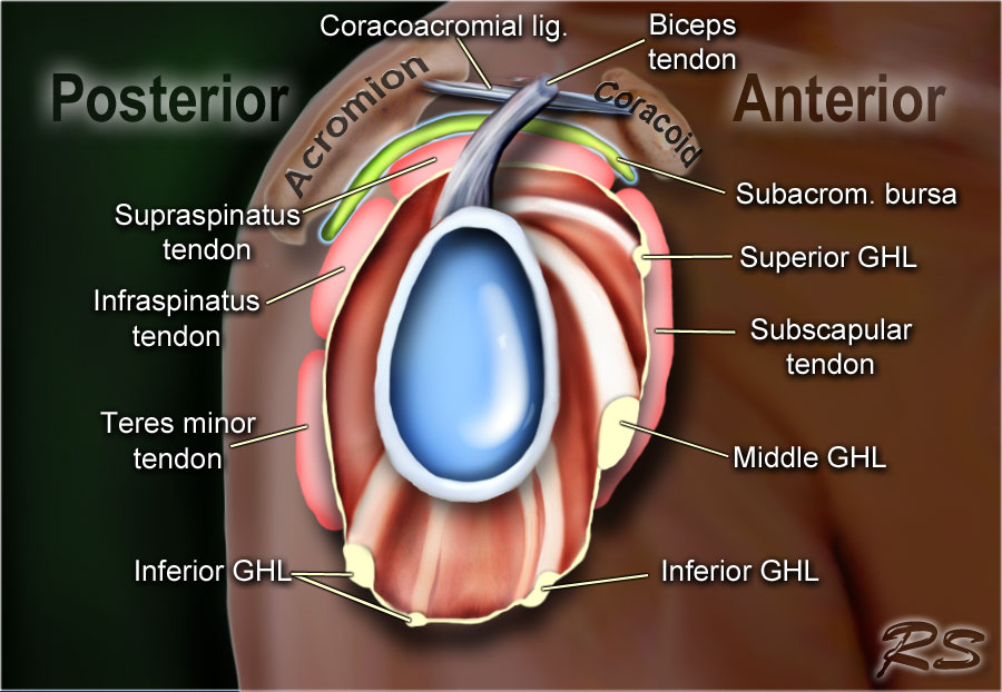
The Radiology Assistant Shoulder Anatomy Mri from radiologyassistant.nl Infraspinatus and teres minor tendon. In this episode of eorthopodtv, orthopaedic surgeon randale c. The subacromial bursa lies on the superior aspect of the supraspinatus tendon (see the images below). Deep to the rtc tendon insertions, blends with the capsule and supraspinatus to form part of the roof of the. The rotator cuff is a group of four muscles and tendons that surround the glenohumeral joint. The shoulder joint (glenohumeral joint) is a ball and socket joint between the scapula and the humerus. More precisely, it consists of several joints that work together. An image depicting shoulder anatomy can be seen below.
The subacromial bursa lies on the top portion of the supraspinatus tendon.
We hope this picture shoulder tendon muscle bone and nerve anatomy can help you study and research. Functional anatomy of the shoulder. Shoulder radiology & anatomy at usuhs.mil. The shoulder joint (glenohumeral joint) is a ball and socket joint between the scapula and the humerus. Learn more about the anatomy the shoulder girdle is the most mobile joint in the human body. Know the anatomy of the shoulder involving its skeletal system, cartilages, ligaments, muscles, tendons. The rotator cuff is a group of four muscles and tendons that surround the glenohumeral joint. The shoulder is a highly complex joint. The shoulder joint is highly mobile and relies on coordination between various muscles, tendons due to its complex anatomy the shoulder is prone to injuries and to degenerative wear and tear such. The long head biceps tendon travels through the shoulder joint making it more prone to injury such as a partial tear, rupture. The nerves supply all the structures above and make them work. In this episode of eorthopodtv, orthopaedic surgeon randale c. More precisely, it consists of several joints that work together.
The shoulder is designed to be incredibly flexible. Notice that the supraspinatus tendon is parallel to the axis of the muscle. Functional anatomy of the shoulder. Normal anatomy, variants and checklist. Just remember the articulating surfaces.
Shoulder Tendon Injury Stock Image C021 1030 Science Photo Library from media.sciencephoto.com The shoulder anatomy provides mobility but leads to a relatively unstable joint, prone to subluxation and dislocation 2. Infraspinatus and teres minor tendon. Look for an os acromiale. An image depicting shoulder anatomy can be seen below. Deep to the rtc tendon insertions, blends with the capsule and supraspinatus to form part of the roof of the. Learn more about the anatomy the shoulder girdle is the most mobile joint in the human body. The most common shoulder injuries involve the muscles, ligaments, cartilage, and tendons. Shoulder radiology & anatomy at usuhs.mil.
The shoulder anatomy provides mobility but leads to a relatively unstable joint, prone to subluxation and dislocation 2.
Your shoulder is made up of three bones: Muscles attachment of rotator cuff muscle | rx harun : Shoulder anatomy is an elegant piece of machinery having the greatest range of motion of any joint in the body. Just remember the articulating surfaces. Shoulder muscles and shoulder tendons. The most common shoulder injuries involve the muscles, ligaments, cartilage, and tendons, rather than the bones. Shoulder tendonitis is inflammation of your rotator cuff or bicep tendons you can develop shoulder tendonitis from participating in certain sports that require the arm to move over the head repeatedly. The shoulder joint is highly mobile and relies on coordination between various muscles, tendons due to its complex anatomy the shoulder is prone to injuries and to degenerative wear and tear such. Your upper arm bone (humerus), your once the ligaments, tendons, and muscles around the shoulder become loose or torn, dislocations can occur. An image depicting shoulder anatomy can be seen below. Sechrest, md narrates an animated tutorial on the basic anatomy of the shoulder. Notice that the supraspinatus tendon is parallel to the axis of the muscle. It is the major joint connecting the upper limb to the trunk.
Infraspinatus and teres minor tendon. Muscles attachment of rotator cuff muscle | rx harun : The shoulder is designed to be incredibly flexible. In addition to shoulder dislocations, other common injuries include rotator cuff tendon tears and broken bones including the humerus and collar terry gc, chopp tm. The shoulder joint is the connection between the chest and the upper extremity.
Alternatives To Rotator Cuff Tear Surgery The Evidence For Non Surgical Options Caring Medical Florida from www.caringmedical.com The tendons and the muscles come next. Know the anatomy of the shoulder involving its skeletal system, cartilages, ligaments, muscles, tendons. Sechrest, md narrates an animated tutorial on the basic anatomy of the shoulder. Home » what is frozen shoulder » shoulder anatomy. This tendon is actually continuous with the glenoid labrum and it runs over the glenohumeral joint and adds a okay! One tendon might have it worse, but it's never isolated to just one tendon. The shoulder joint (glenohumeral joint) is a ball and socket joint between the scapula and the humerus. Your shoulder is made up of three bones:
The subacromial bursa lies on the top portion of the supraspinatus tendon.
Subscapular bursa or the scapulothoracic bursa: In addition to shoulder dislocations, other common injuries include rotator cuff tendon tears and broken bones including the humerus and collar terry gc, chopp tm. Shoulder anatomy is a remarkable combination of strong bones, flexible ligaments and tendons, and reinforcing cartilage and muscles. Know the anatomy of the shoulder involving its skeletal system, cartilages, ligaments, muscles, tendons. Shoulder tendonitis is inflammation of your rotator cuff or bicep tendons you can develop shoulder tendonitis from participating in certain sports that require the arm to move over the head repeatedly. It is the major joint connecting the upper limb to the trunk. The subacromial bursa lies on the superior aspect of the supraspinatus tendon (see the images below). Home » what is frozen shoulder » shoulder anatomy. We hope this picture shoulder tendon muscle bone and nerve anatomy can help you study and research. The long head biceps tendon travels through the shoulder joint making it more prone to injury such as a partial tear, rupture. Look for an os acromiale. The nerves supply all the structures above and make them work. The rotator cuff is a group of four muscles and tendons that surround the glenohumeral joint.


0 Komentar